Translate this page into:
Pregnancy and Unilateral Krukenberg Tumour: Factual or Fictious – A Case Report

*Corresponding author: Dr. Joel Abraham Thomas, Department of Radiation Oncology, Christian Medical College, Ludhiana, Punjab, India. joelexams@gmail.com
-
Received: ,
Accepted: ,
How to cite this article: Thomas JA, Mathew RM, Angela DD, Chauhan H, Kingsley PA. Pregnancy and Unilateral Krukenberg Tumour: Factual or Fictious – A Case Report. Indian Cancer Awareness J. 2024;3:17-21. doi: 10.25259/ICAJ_14_2024
Abstract
Krukenburg tumor, a secondary ovarian neoplasm, arises from various primary sites. It’s occurrence during pregnancy is extremely rare. We are reporting the case of a 24 year old G2P1L1 lady with a unilateral Krukenberg tumor diagnosed in first trimester of pregnancy. The case posed as a diagnostic challenge as the presentation is non-specific and radiological diagnosis is very challenging owing to the similarity of the tumour to ectopic pregnancy on imaging. The sparsity of data and unique presentation of the patient inspired us to write this report. This is a 24 year old lady who presented to the emergency department with the complaints of amenorrhea for 2 months, pain abdomen and bleeding per vaginum for 2 days. Urine Pregnancy Test done at home was positive. She was found to be in shock and was resuscitated. Ultrasonography done showed a possibility of an ectopic pregnancy with rupture or a partial molar pregnancy. She was taken up for emergency laparotomy, a lobulated, irregular right sided ovarian mass suggestive of a right ovarian neoplasm with a normal left ovary and an intrauterine incomplete abortion was found. She underwent right salpingo-oophorectomy along with the ovarian mass; with stepwise exploration of pelvis and abdomen followed by dilatation and curettage for the evacuation of the retained product of conception. Histopathological examination revealed features suggestive of metastatic Signet Ring Cell Carcinoma. Further investigation with CECT chest, abdomen and pelvis and upper GI endoscopy revealed a gastric primary. This case depicts the challenges faced by physicians in correctly diagnosing malignant tumours during pregnancy. By reporting this case, we aim to raise awareness among physicians regarding the need for heightened vigilance and multi-disciplinary approach in order to timely and accurately diagnose such conditions.
Keywords
Krukenberg
Pregnancy
Unilateral
INTRODUCTION
During pregnancy, the occurrence of adnexal masses ranges from 1 in 81 to 1 in 2500 pregnancies. Among these masses, only 3% are found to be malignant.[1] The occurrence of a Krukenberg tumour (KT) originating from gastric cancer during pregnancy is even more uncommon. Notably, gastric cancer, particularly in young women, is susceptible to ovarian metastasis.[2] The German pathologist Friedrich Krukenberg was the first to report gastric cancer with ovarian metastasis in 1896. He named the tumour after himself, calling it KT.[3] The term “Krukenberg tumour” refers to a malignancy found in the ovary that originates from a primary site elsewhere in the body, typically the gastrointestinal (GI) tract. In the majority of cases, KT arises from a primary tumour of the GI tract, usually originating from the stomach and colorectum.[4] Often, these tumours are bilateral (over 80%), given their metastatic nature.[5] Novak and Gray identified the diagnostic criteria for KT and included the presence of an ovarian neoplasm along with signet ring cells producing mucin and sarcomatoid proliferation of the ovarian stroma.[6]
Ovarian metastasis occurs in approximately 5–10% of female gastric cancer cases and has been reported in 33–41% of autopsies.[7] Pregnancy complicated by KTs is exceptionally rare; however, these tumours can present with non-specific GI signs and symptoms such as nausea, vomiting, abdominal or pelvic pain, bloating and ascites. These symptoms may be difficult to discern during pregnancy, causing challenges in diagnosis and leading to poor prognosis and outcome.
We present a rare case of gastric carcinoma diagnosed during the first trimester of pregnancy. This is the first reported case of a unilateral KT diagnosed during pregnancy, which caused a diagnostic dilemma as it clinically mimicked an ectopic pregnancy with the potential for rupture and a partial molar pregnancy. By reporting this case, we aim to raise awareness among physicians regarding the need for increased vigilance when encountering abnormal adnexal masses accompanied by vaginal bleeding and abdominal pain in pregnant women.
CASE REPORT
An un-booked 24-year-old (Gravida – 2, Para– 1, Living – 1, Neonatal Death - 1) Indian lady, a chronic smokeless tobacco chewer, presented to the emergency department with the complaints of amenorrhea for 2 months, pain in abdomen and bleeding per vaginum for 2 days. A urine pregnancy test done at home was positive. She was evaluated in a peripheral hospital for the aforementioned complaints, and her ultrasonogram (USG) abdomen showed features suspicious of a ruptured ectopic pregnancy, and thereby, she was referred to our centre. On arrival at the emergency department, she was evaluated and found to have severe blood loss anaemia, which caused compensated hypovolemic shock. She was resuscitated with IV crystalloids and cross-matched blood transfusions. USG showed a large hyperechoic mass ~ 14.8 cm (AP) × 13.2 cm (TR) with few cystic spaces within, arising from the right adnexa showing significant internal vascularity – the possibility of ectopic pregnancy with rupture or a partial molar pregnancy was suspected. Mild-to-moderate free fluid in the abdomen and pelvis with few fine free-floating internal echoes within was noted – likely hemoperitoneum. Heteroechoic contents did not show vascularity on colour. Doppler imaging was seen within the cervical canal, likely blood clots. A bulky uterus (~7.0 × 4.1 cm) with a thickened endometrial stripe (14 mm) and cystic structure (1.0 × 0.4 cm) within the lower endometrial cavity was visualised. Considering her haemorrhagic hypovolemic shock status, she was taken up for emergency laparotomy under general anaesthesia. Intraoperatively, there was a 20 × 14 × 8 cm lobulated, irregular right-sided ovarian mass, which weighed approximately 1000 mg, suggestive of a right ovarian neoplasm with a normal left ovary and an intrauterine incomplete abortion [Figure 1]. She underwent right salpingo-oophorectomy along with the ovarian mass, with a stepwise exploration of the pelvis and abdomen, followed by dilatation and curettage for the evacuation of the retained product of conception. She tolerated the procedure well and her post-operative period was uneventful.

- (a) and (b) Intraoperative finding showing right ovarian mass and normal left ovary (blue arrow), (c) The dissected right ovarian mass.
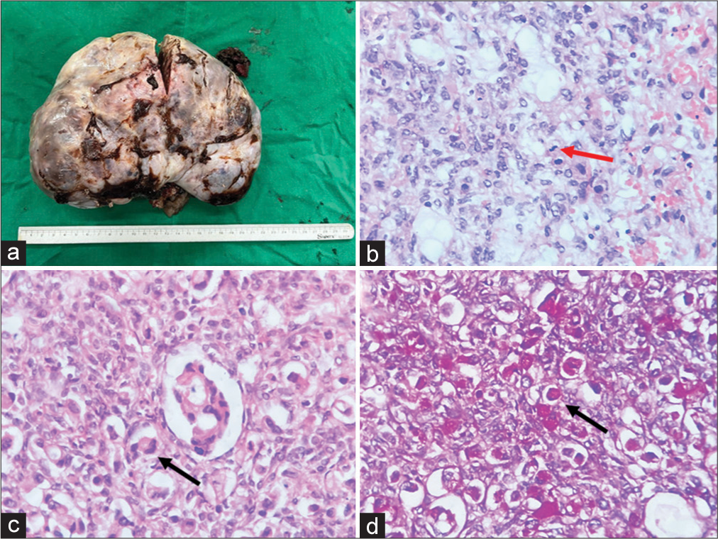
Histopathological examination revealed features suggestive of metastatic signet ring cell carcinoma to the right ovary [Figure 2]. Cancer antigen (CA) 125 levels were elevated to 629, CA 19/9–220, and carcinoembryonic antigen–14.90. Lactate dehydrogenase was 368, and beta-human chorionic gonadotropin was 946.9.

- Right ovarian Kruckenberg tumour. (a) Gross image showing a large lobulated ovarian mass weighing 1,100 gm. (b) Histopathology (hematoxylin and eosin) showing occasional glandular lumina formation, increased mitotic activity (red arrow) and abundant intra-cytoplasmic mucin, (c) Histopathology (hematoxyllin and eosin) showing large signet ring cells (black arrow), (d) Diastase-resistant periodic acid Schiff (DPAS) stain showing large signet ring cells (black arrow).
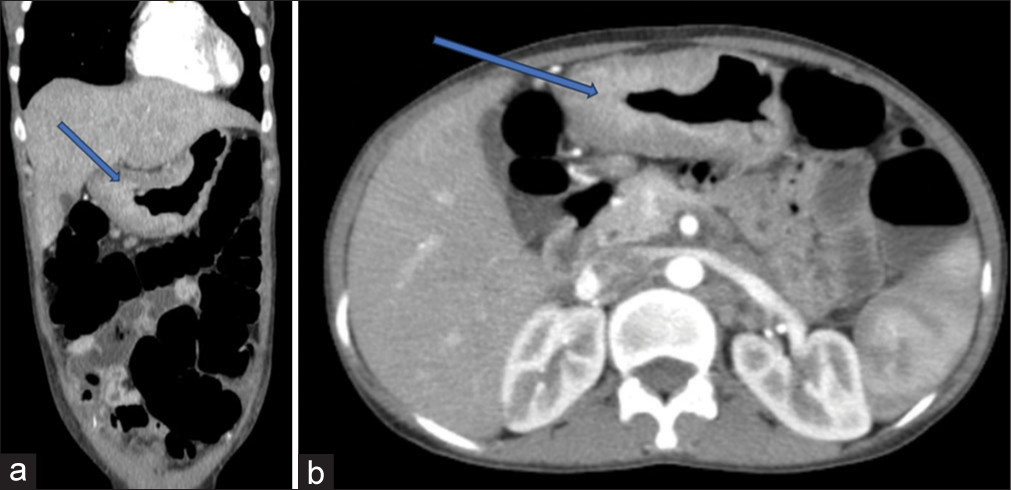
Keeping the possibility of a GI primary, she underwent contrast-enhanced computerised tomography chest, abdomen and pelvis which showed irregular, nodular, circumferential, asymmetrical heterogeneously enhancing wall thickening in the stomach involving distal body, pylorus, antrum and pyloro-duodenal junction having maximum thickness up to 25 mm in the pylorus causing moderate luminal narrowing at the pylorus along with abdominal lymphadenopathy [Figure 3].

- Contrast enhanced computerised tomography (CECT) images showing the stomach lesion involving distal body, pylorus, antrum and pyloro-duodenal junction (blue arrows) a) coronal view. b) CECT axial view..
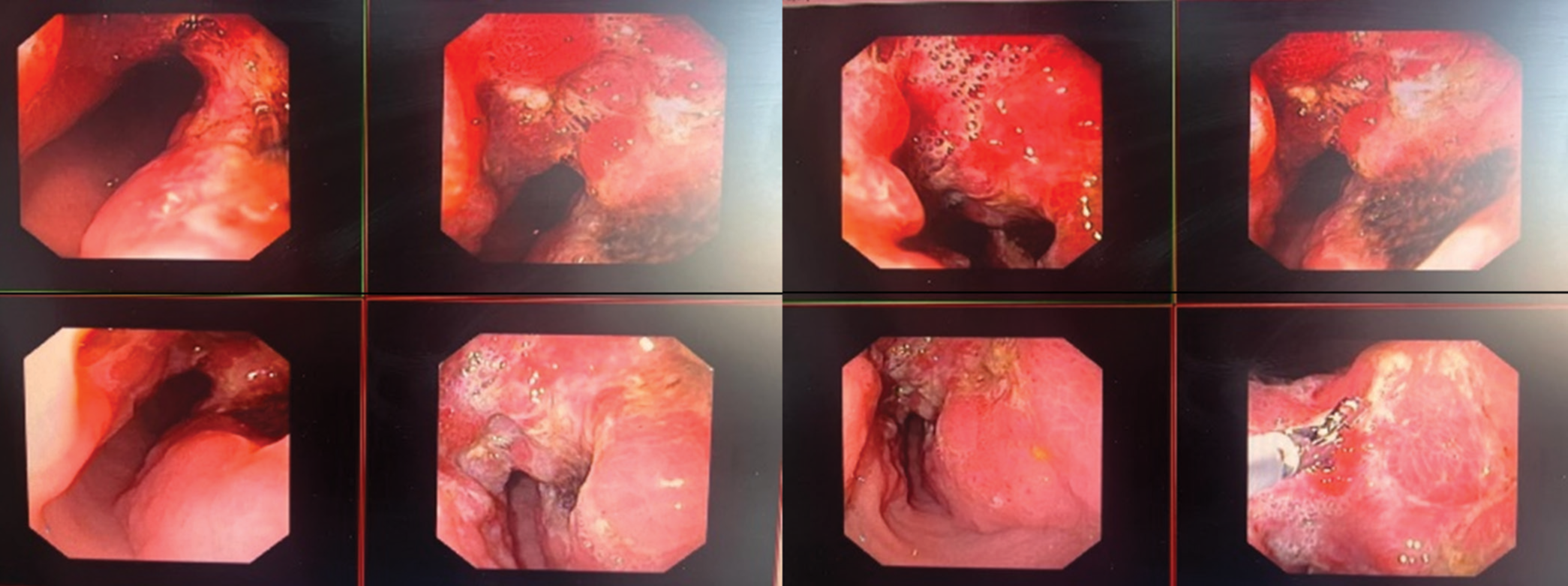
Upper GI endoscopy showed that a large irregular ulcer with elevated margins was seen in the distal body and antrum, along with the lesser curvature of the stomach [Figure 4].

- Upper gastrointestinal endoscopic images showing the gastric lesion at distal body and antrum, along with the lesser curvature of the stomach.
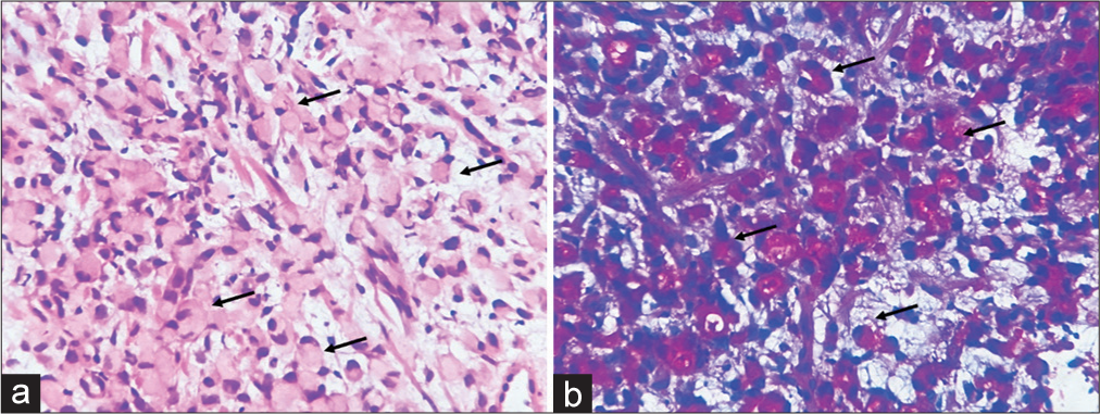
A biopsy was taken from the gastric lesion, and when evaluated histopathologically, it revealed features suggestive of signet ring cell carcinoma of the stomach with Her2ƞ Positive, Score 3+ [Figure 5].

- Gastric biopsy showing signet ring cell carcinoma. (a) High-power view showing numerous signet ring cells (black arrows), ×400; hematoxylin and eosin and (b) signet ring cells filled with intra-cytoplasmic mucin (black arrows), ×400; combined Alcian blue-Periodic acid Schiff stain.
DISCUSSION
The emergence of a KT stemming from gastric carcinoma during pregnancy is exceptionally rare. Typically, gastric carcinoma manifests in older demographics, predominantly males, but exhibits a higher incidence among younger females in this scenario.[8] According to the Global Cancer Statistics for 2020, gastric cancer stands as the fifth most common cancer by incidence and the fourth highest in terms of mortality among all malignancies worldwide.[9] Metastasis represents a notable feature of gastric cancer advancement and stands as the primary factor contributing to mortality linked with the illness.
The case of unilateral ovarian KT in a pregnant woman presents a unique clinical scenario with significant diagnostic and therapeutic challenges. Krukenberg tumour, a secondary ovarian neoplasm, arises from various primary sites, including the stomach, colon, rectum, breast, oesophagus, gallbladder and appendix. Gastric carcinoma, comprising roughly 70% of cases, stands as the most frequent primary source. The median overall survival for individuals with Krukenberg tumors stemming from gastric origins is noted to be 11 months.[10] In the context of pregnancy, the diagnosis and management of such cases become even more complex due to the overlapping clinical presentations with common obstetric conditions, and the imperative is to safeguard both maternal and foetal health. However, gastric cancer diagnosed during pregnancy is commonly detected at advanced stages, resulting in a bleak prognosis for both the mother and the foetus. This discussion will delve into the clinical implications, diagnostic considerations, management strategies and prognostic factors associated with this rare manifestation of metastatic gastric cancer.
The clinical presentation with amenorrhea, abdominal pain and vaginal bleeding could be misconstrued as signs of various obstetric complications, including threatened or spontaneous abortion, ectopic pregnancy and molar pregnancies. Ultrasonography plays a pivotal role in evaluating adnexal masses; however, its accuracy in distinguishing between benign and malignant masses is limited. The initial suspicion of a ruptured ectopic pregnancy based on ultrasound findings highlights the diagnostic dilemma of distinguishing between obstetric emergencies and rare oncological conditions occurring during pregnancy. Ultrasonography serves as the primary imaging modality; however, its limitations in distinguishing between benign and malignant masses necessitate further investigations, such as computed tomography or magnetic resonance imaging. In this case, the presence of a large heteroechoic mass arising from the right adnexa with internal vascularity was suggestive of an ovarian pathology, prompting further investigation.
The occurrence of a KT during pregnancy is exceedingly rare and is further compounded by its unilateral presentation. The diagnostic dilemma is heightened by the overlapping symptoms with obstetric conditions such as ectopic pregnancy or molar pregnancy. Moreover, the aggressive nature of KTs, particularly in the context of metastatic gastric cancer, underscores the urgency for accurate diagnosis and prompt intervention to optimise maternal and foetal outcomes. The diagnosis of KT relies on a combination of clinical presentation, radiological imaging and histopathological examination. The definitive diagnosis of KT is established through histopathological examination, characterised by the presence of signet ring cells producing mucin and sarcomatoid proliferation of the ovarian stroma.
CONCLUSION
This case of a unilateral ovarian KT in pregnancy highlights the complexity of diagnosing and managing rare oncological conditions during gestation. Timely recognition, accurate diagnosis and multidisciplinary collaboration are imperative to optimise maternal and foetal outcomes in such cases. The 5-year survival rate for women affected by gastric cancer is documented as zero, with many instances culminating in death within 6 months following diagnosis. Consequently, the prognosis for this condition tends to be grim. The median overall survival for individuals with KTs stemming from gastric origins is noted to be 11 months. By reporting this case, we aim to raise awareness among clinicians regarding the need for heightened vigilance while encountering adnexal masses in pregnant women, particularly in the context of non-specific GI symptoms. Further, research is warranted to elucidate optimal management strategies and prognostic indicators in this unique patient population.
Acknowledgement
1) Dr. Pamela Alice Kingsley, Professor and Head, Department of Radiation Oncology
2) Dr. Preethy Mercy Paul, Professor and Head, Department of Pathology
3) Dr. Harpal Singh, Associate Professor and Head, Department of Gastroenterology
4) Dr. Kavita Bhatti, Professor and Head, Department of Obstetrics and Gynecology.
Ethical approval
The Institutional Review Board approval is not required.
Declaration of patient consent
The authors certify that they have obtained all appropriate patient consent.
Conflicts of interest
There are no conflicts of interest.
Use of artificial intelligence (AI)-assisted technology for manuscript preparation
The authors confirm that there was no use of artificial intelligence (AI)-assisted technology for assisting in the writing or editing of the manuscript and no images were manipulated using AI.
Financial support and sponsorship
Nil.
References
- Gastric Cancer During Pregnancy: A Report on 13 Cases and Review of the Literature with Focus on Chemotherapy During Pregnancy. Acta Obstet Gynecol Scand. 2020;99:79-88.
- [CrossRef] [PubMed] [Google Scholar]
- Cancer Statistics in China, 2015. CA Cancer J Clin. 2016;66:115-32.
- [CrossRef] [PubMed] [Google Scholar]
- An in-depth look at Krukenberg tumor: An overview. Arch Pathol Lab Med. 2006;130:1725-30.
- [CrossRef] [PubMed] [Google Scholar]
- An In-depth Look at Krukenberg Tumor: An Overview. Arch Path Lab. 2006;130:1725-30.
- [CrossRef] [PubMed] [Google Scholar]
- Adenocarcinoma of Sigmoid Colon with Metastasis to an Ovarian Mature Teratoma: A Case Report. World J Clin Cases. 2024;12:853-8.
- [CrossRef] [PubMed] [Google Scholar]
- Krukenberg Tumor of the Ovary: Clinical and Pathological Study of Four Cases. Surg Gynecol Obstet. 1938;66:157-65.
- [Google Scholar]
- Prognostic Factors for Ovarian Metastases from Primary Gastric Cancer. Int J Gynecol Cancer. 2008;18:825-32.
- [CrossRef] [PubMed] [Google Scholar]
- Trends in Gastric Cancer Sex Ratio in the United States. Cancer. 1987;59:1032-5.
- [CrossRef] [PubMed] [Google Scholar]
- Global Cancer Statistics 2020: GLOBOCAN Estimates of Incidence and Mortality Worldwide for 36 Cancers in 185 Countries. CA Cancer J Clin. 2021;71:209-49.
- [CrossRef] [PubMed] [Google Scholar]
- Krukenberg Tumor of Gastric Origin in Pregnant Women with Preeclampsia. Case Rep Oncol. 2023;16:712-21.
- [CrossRef] [PubMed] [Google Scholar]


